Last Updated: January 14, 2025
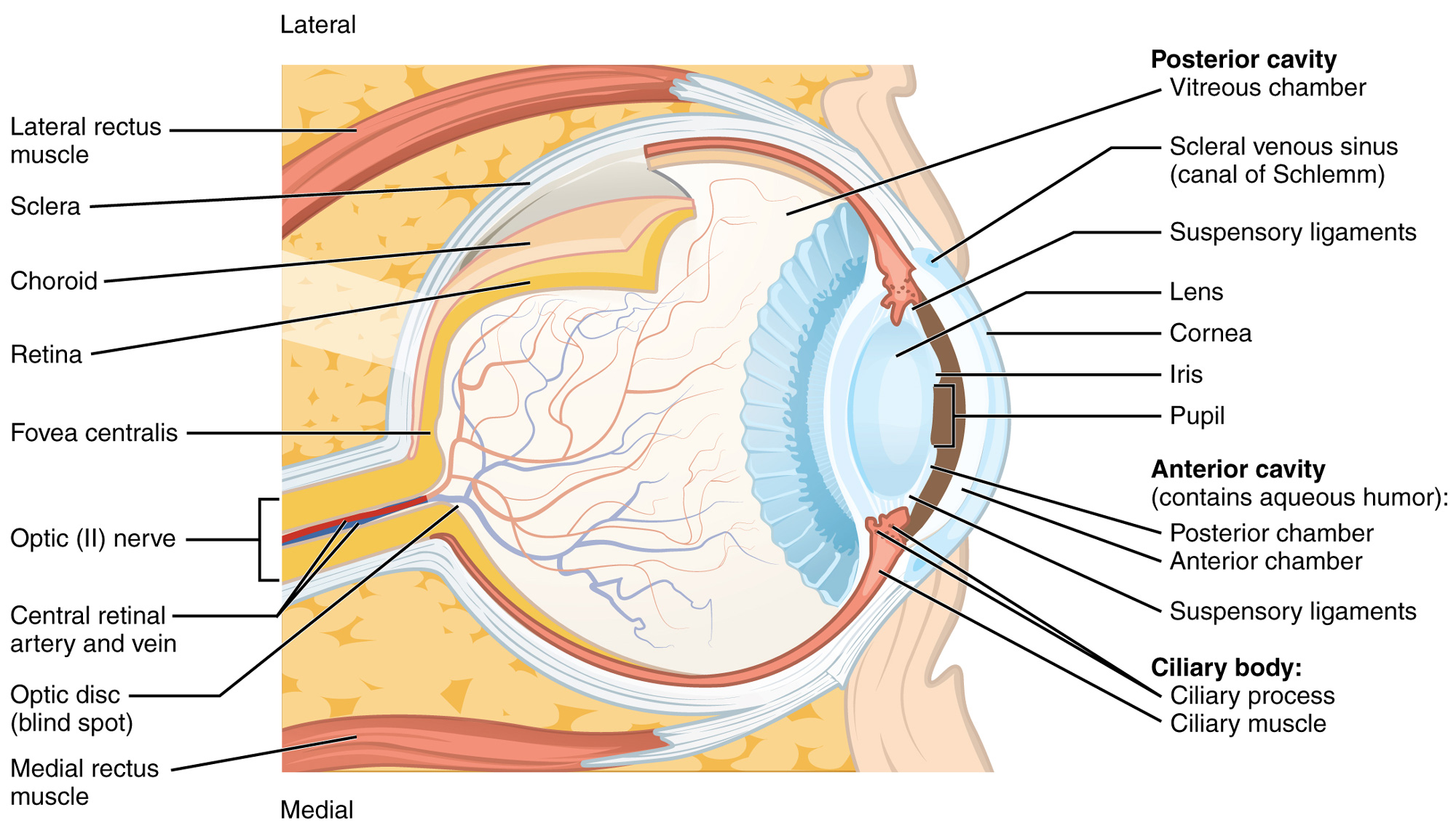
Structure of the eye. Source: OpenStax College via Wikimedia Commons
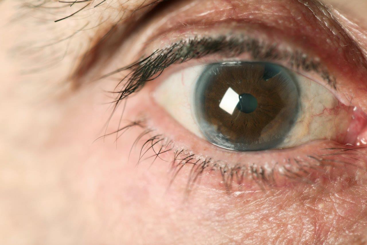
The front surface of the eye is called the cornea, a transparent layer that offers protection and allows for light to enter the eye. Surrounding the cornea is the white parts of the eye called the sclera, which wraps around and provides the round structure of the eyeball. Behind the cornea is a fluid-filled space called the anterior chamber and behind this space is the coloured part of the eye called the iris. The iris is the most distinct feature that we notice when we look at someone eyes. Different eye colours exist due to a difference in melanocytes in the irises, the same cells responsible for the production of our skin colour. In the middle of the iris is a hole called the pupil, which can widen or narrow as the iris around it contracts or relaxes. The pupil controls the amount of light that enters into the eye. Behind the pupil and the iris is another fluid-filled space called the posterior chamber. The fluid that fills the anterior and posterior chambers is called aqueous humor which is produced by the ciliary body. If you’ve ever wondered what the front of the human eye looks like at high resolution, photographer Suren Manvelyan has captured it through his series “Your Beautiful Eyes”.
Behind the posterior chamber is a transparent disk called the crystalline lens, which serves to further transmit and focus the incoming light towards the back of the eye. The lens is held in place by string-like suspensory ligaments called zonules. When the lens turns cloudy or opaque, it blocks the incoming light and decreases our ability to see, this is called a cataract.
Behind the lens is a large space taking up most of the volume of the eye, called the vitreous body which is filled with a jelly-like substance called the vitreous humor. Behind the vitreous body, at the very back of the eye, is a layer called the retina, which contains millions of light-sensitive cells that detects light. These detected light signals all gather at a structure called the optic nerve on the retina, which connects the eye to the brain. This is how we are able to perceive the world when we look at something. Throughout the retina, blood vessels are present to supply nourishment to this tissue. Located near the center of the retina is a structure called the macula, this area provides clear and sharp central vision important for our daily tasks, such as reading.
Other structures surrounding the eye that help with its function include the eyelids which is a thin fold of skin that covers and protects the front of the eye, and extraocular muscles that attach to the eyeball to allow for eye movements.
To learn more about your eyes and to ensure they are healthy, book an eye examination with your local optometrist.
References
- Oyster CW. (1999). The human eye. Sunderland, MA: Sinauer.
- Image: Anatomy & Physiology, OpenStax College via Wikimedia Commons, Accessed June 9th 2022.
Related Articles